|
|
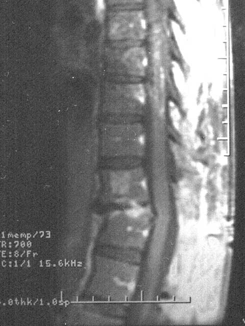
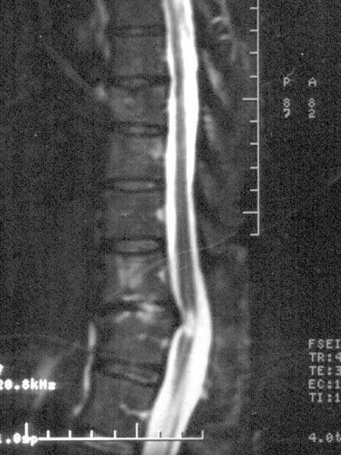
Magnetic Resonance Imaging (MRI) Cross Section: |
|
|
Lack of Disc Support |
Pinched Spinal Cord |
Pause mouse over a picture to pull up an alternate description. Click on a picture to enlarge it.
A bony structure (the vertebral, or spinal, column) protects the spinal cord, an extension of the brain. All information from the brain to the arms and legs travels through the spinal cord, and allows movement. The center of the back involves the thoracic region. A disc reacts as a spongy shock absorber to cushion and separate the bones (vertebrae) of the spine. Ruptured disc damage—one vertebra gnashing against an adjacent bone, pinching and crunching spinal nerves that are located behind the discs, possibly with intense pain—and pressure on the spinal cord severe and long lasting can cause a total and permanent loss of function below the level of pressure (from Ruth's waist, down).
Dr. Hipp, October 4, 2004: Ruth is continuing to make improvements. MRI and return appointment in 6 months.
Also, both Ruth & Joe are personally grateful to 2003's Nobel Prize medicine winners, Briton Peter Mansfield & American Paul Lauterbur, whose discoveries led to MRI development, enabling 3-D details showing up on Ruth's MRI's taken November 20, 2003 & October 1, 2004.
Our Home Page Our Families Page Ruth's Disc Rupture & Recuperation Record